


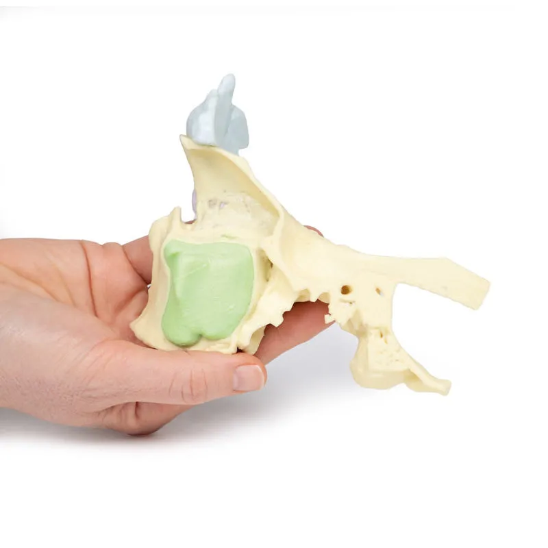
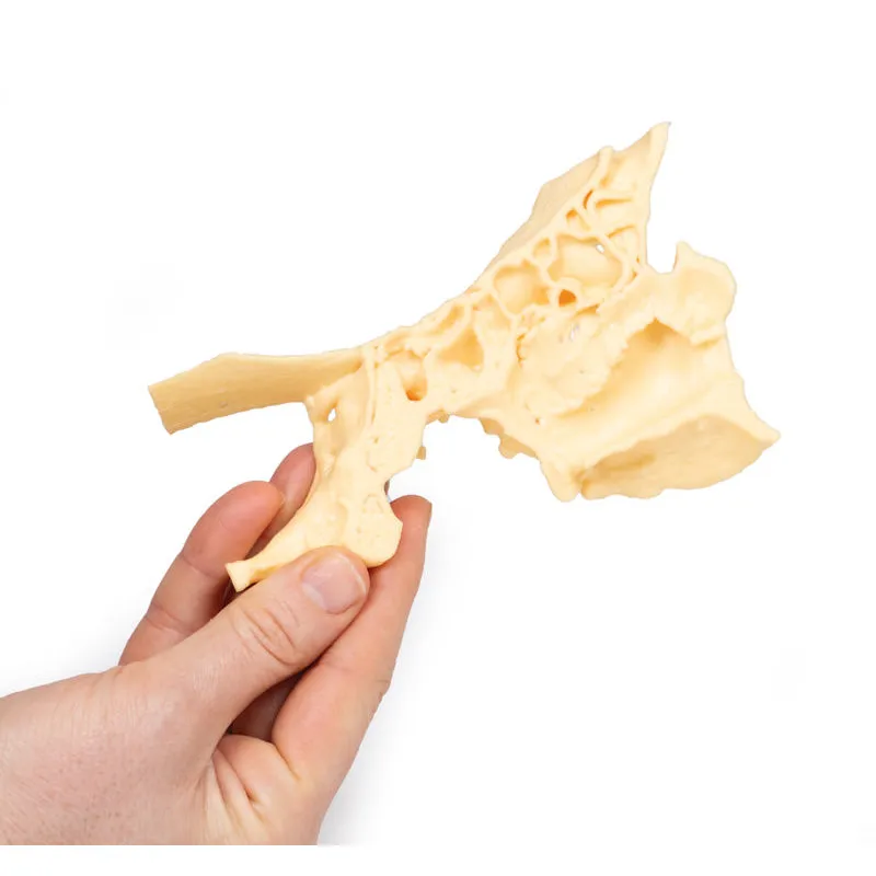
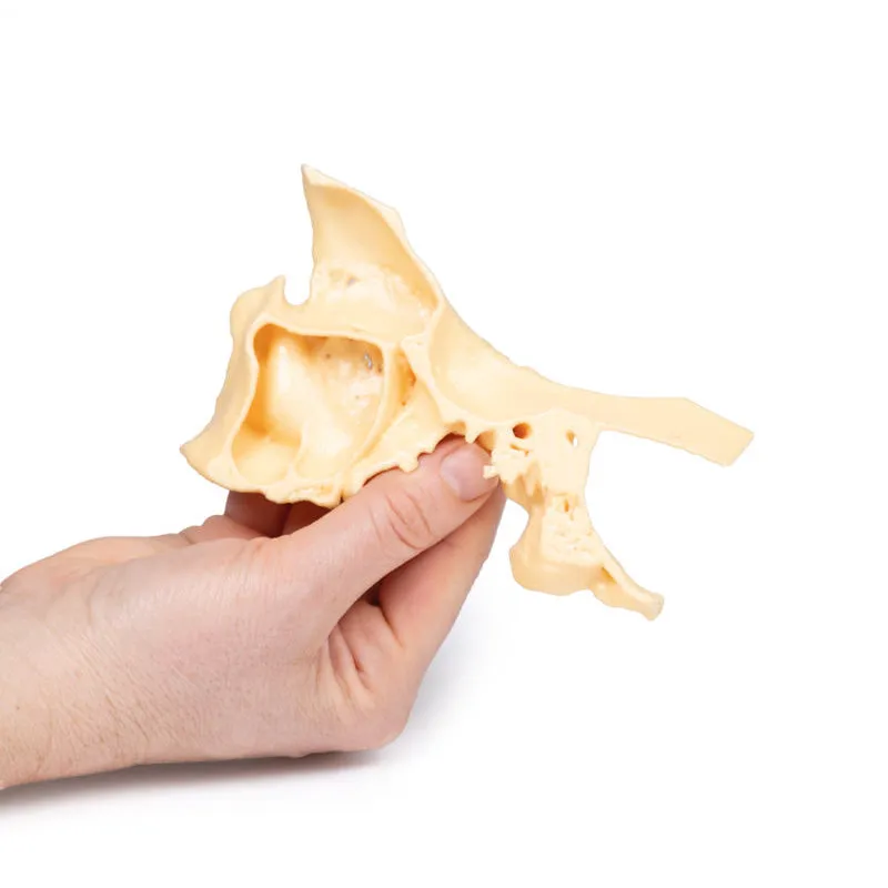
3D Printed Paranasal Sinus Model
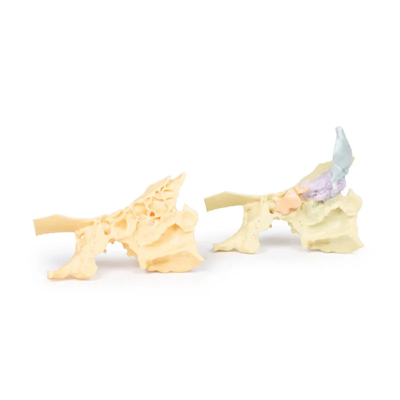
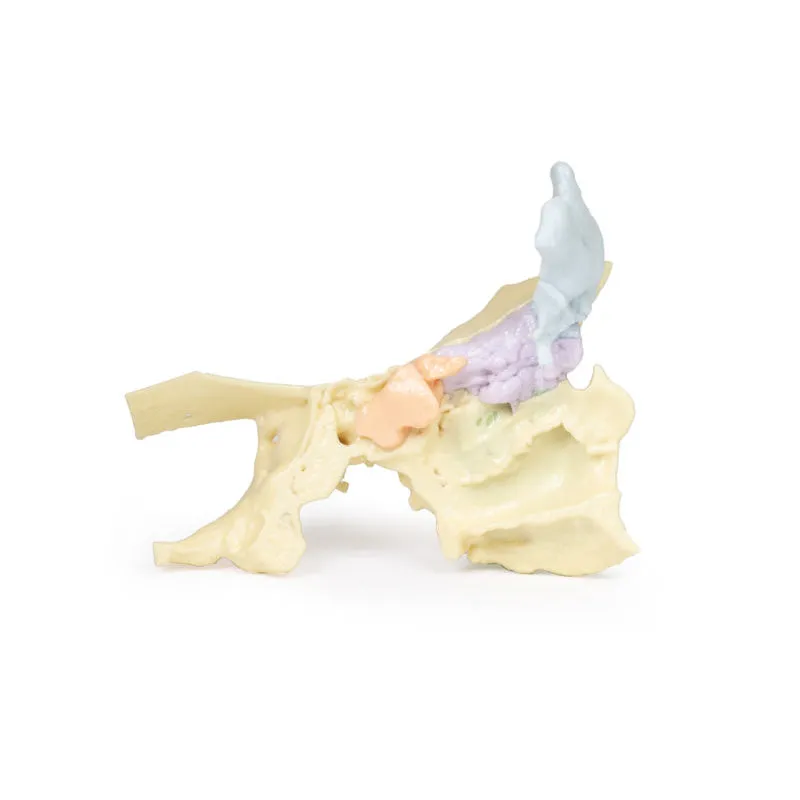
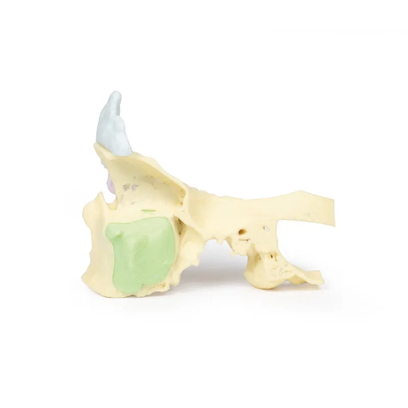
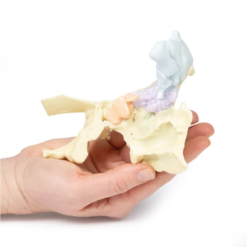
This unique model has been created from CT imaging and segmentation of the internal spaces of the viscerocranium. Parts of the skull have been retained but sections or windows have been removed to expose the paranasal sinuses. The paired frontal sinuses, with the right being partially subdivided, are coloured blue. The left frontal sinus is largely surrounded by frontal bone, while the right is completely exposed and shows the frontonasal ostium which drain as a funnel shaped tube into the infundibuklum of the middle meatus of the nasal cavity. The ethmoid sinuses or air cells, coloured purple, are only shown on the left.The medial wall of the orbit composed of the orbital plate of the ethmoid bone is retained. The maxillary sinus (green) on the left has been partly exposed and partly left within the maxilla. The opening of the maxillary sinus into the lateral wall of the nose is barely discernable as a small green patch in the middle meatus. The left sphenoid sinus (pink) is also displayed, within the opened sphenoid bone.
GTSimulators by Global Technologies
Erler Zimmer Authorized Dealer


")
