


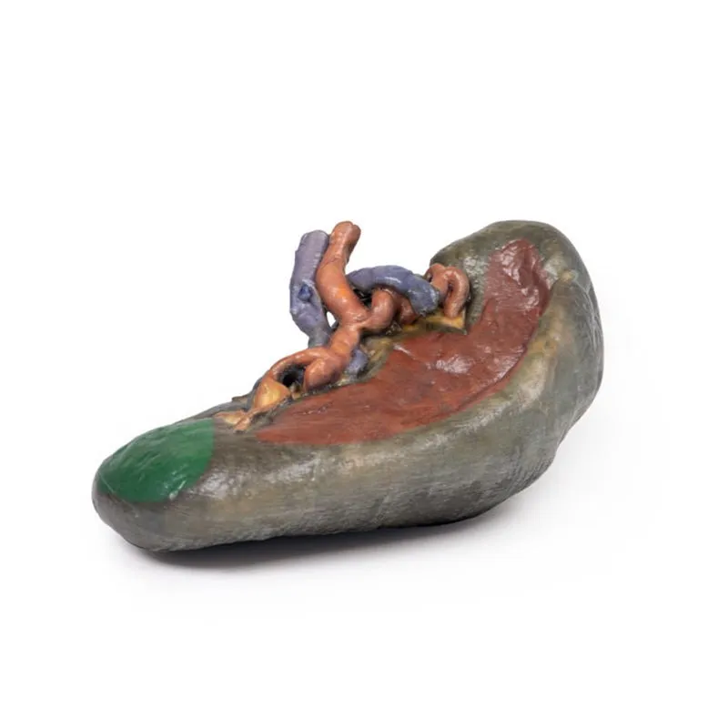
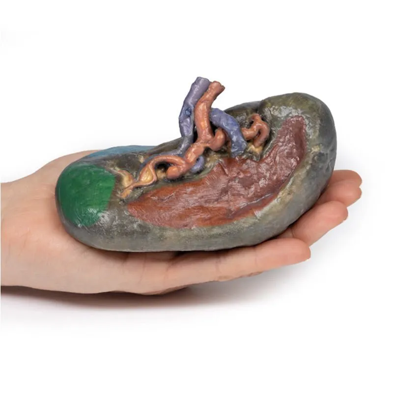
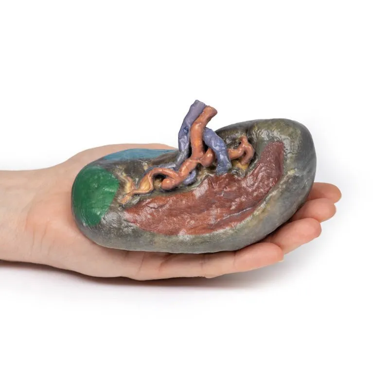
3D Printed Vasculature of the Spleen
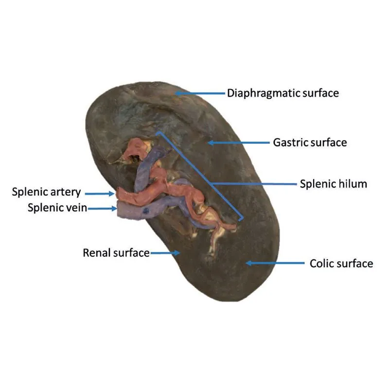
At the splenic hilum, the splenic artery and vein can be seen entering the spleen to
supply and drain the organ. The opening of the splenic vein has been kept patent by the insertion of silicon tubing
in the model. This model shows the most superior branch of the splenic vein has been sectioned from its normal
passage into the spleen. The “tortuous” of twisted shape of the splenic artery can be appreciated as it branches at
the hilum. This reflects the overall curled and twisted shape of the vessel across its course from the coeliac trunk
to the spleen.
The splenic artery and vein give rise to short gastric vasculature as well as the left gastro-omental vasculature.
In this specimen, the splenic artery and vein have been cut after these vessels have branched, and thus they cannot
be seen in the model.
The splenorenal ligament connects the spleen to the left kidney and contains the splenic artery, splenic vein and
tail of the pancreas. It is formed by the overlaying of peritoneum that was originally part of the dorsal mesentery
during embryological development over this vasculature. The splenorenal ligament is not observable on the model as
the peritoneum has been removed in order to expose the splenic vasculature.
The gastrosplenic ligament connects the stomach to the spleen and contain the short gastric arteries and part of the
left gastro-omental artery at its origin as it branches off the splenic artery. Like the splenorenal, the
gastrosplenic ligament is formed by the overlaying of peritoneum that was originally part of the dorsal mesentery
during embryological development. The gastrosplenic ligament is not present as the splenic artery has been dissected
after its formation.
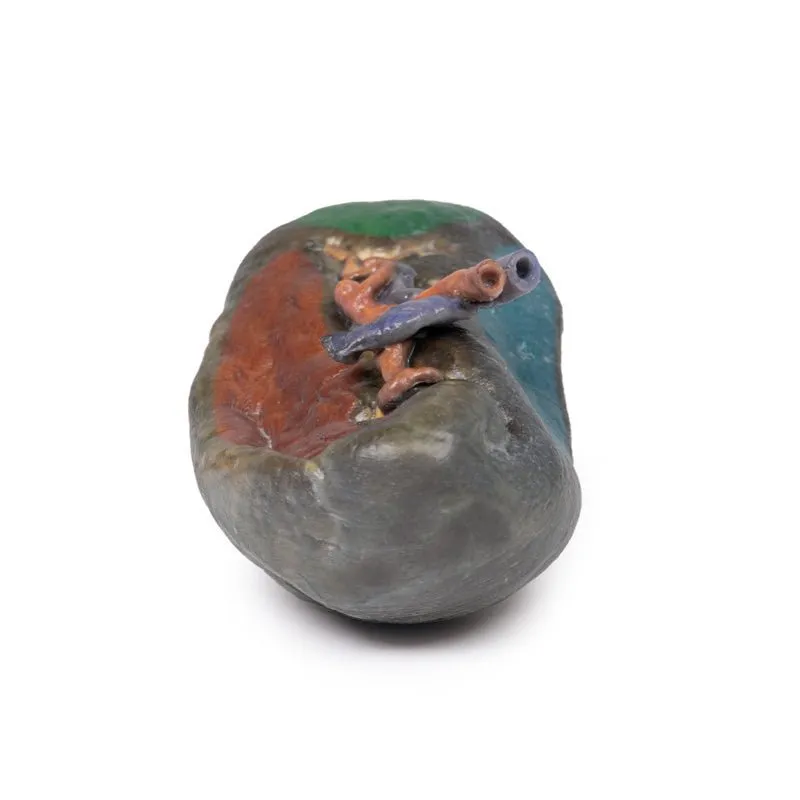
The outside of the spleen consists of a thin fibrous capsule. Due to its delicate nature and the large quantity of
blood usually contained in the spleen, the fibrous capsule is vulnerable to rupture.
GTSimulators by Global Technologies
Erler Zimmer Authorized Dealer


